Services

No category

Acne Scarring

Acne Scarring

Acne scars are usually the result of inflamed blemishes caused by skin pores engorged with excess oil, dead skin cells and bacteria. The pore swells, causing a break in the follicle wall. Shallow lesions are usually minor and heal quickly. But if there is a deep break in the wall of the pore, infected material can spill out into surrounding tissue, creating deeper lesions. The skin attempts to repair these lesions by forming new collagen fibers. These repairs usually aren’t as smooth and flawless as the original skin. There are different types of acne scars. Ice pick scars are flat, pitted scars. Atrophic scars are thin and flat, and often appear depressed when compared to normal surrounding skin. Finally, hypertrophic or keloidal scars are typically firm and raised. Acne scars may be treated with microdermabrasion, soft tissue fillers, laser treatments, and chemical peels.

Actinic Keratoses

Actinic Keratoses

Actinic keratoses (AKs) are precancerous skin lesions that if left untreated may evolve into squamous cell carcinomas. Typically, they occur in fair skinned individuals after the skin has been badly damaged by ultraviolet (UV) rays from the sun or indoor tanning. Actinic keratoses are sandpaper-like rough, red spots. Patients feel them before they see them. Most often, they are found on the skin that is sun exposed: the forehead, ears, neck, arms,
hands, lower lip and bald scalps.

Prevention of actinic keratoses begins with avoiding prolonged sun exposure and using sunscreen. These measures should start in childhood, as the sun damage responsible begins in early life.

There are a number of treatment options available. Cryosurgery, or freezing of involved areas with a cold spray made of liquid nitrogen, is first line for patients with few lesions. For widespread actinic keratoses, field therapy is the treatment of choice and includes topical medications (chemotherapeutics, immunomodulators and other agents) applied at home, as well as in-office procedures using lasers, photodynamic therapy and chemical peels.

Age Spots

Age Spots

As people age, unsightly blemishes, commonly called age spots, can appear on the face and on the back of the hands. The spots, also called lentigines, lentigos or liver spots, are sharply defined, rounded, brown or black, flat patches of skin.

Age spots occur when the top surface layer of skin expands with more pigment and develops what looks like a large freckle. One may appear by itself, or a few may be clustered together.

Some people have a hereditary predisposition to age spots. Age spots may develop at an early age, even in childhood, though they are more common in older individuals, especially in those who have too much sun exposure.

Age spots are not cancerous, nor do they lead to cancer. However, on skin exposed to the sun, they may be accompanied by precancerous scaly, red elevations called actinic keratoses. Lentigenes may be treated with Intense Pulse Light (IPL), laser, chemical peels, and cryosurgery.

Alopecia Areata

Alopecia Areata

Alopecia Areata is a condition in which patients develop small round patches of hair loss. These patches usually grow back on their own or may persist for years. It is most common in people with a family history of the disease.

In alopecia areata, the body’s immune system attacks the root of the hair for unknown reasons. While most people get one or two small patches on their head, a rare minority of people can lose all of their scalp (alopecia totalis) and body hair (alopecia universalis).

Nails may develop tiny pinpoint dents. Alopecia Areata is not associated with any systemic health issues, but people with the disease may be more prone to allergies, asthma, eczema, thyroid dysfunction and vitiligo.

Alopecia Areata usually gets better by itself, but the timing is unpredictable. While there is no cure, the most common treatment is injection of corticosteroids into the affected areas to speed hair recovery.

Angiomas

Angiomas

Angiomas are benign growths that clinically present themselves on the surface of the skin as red bumps or papules. They are derived from cells of the vascular or lymphatic vessel walls (endothelium)

Angiomas are a frequent occurrence as patients age, and they might be an indicator of systemic problems such as liver disease. They are not commonly associated with malignancy. When they are removed, it is generally for cosmetic reasons. They may be removed by laser or a small surgical procedure.

Atypical (Dysplastic) Nevus

Atypical (Dysplastic) Nevus

An atypical or dysplastic nevus is a non-cancerous mole that shares some features with melanoma, but is completely benign. While individual dysplastic nevi do not usually become melanomas, they may be markers for an increased risk of melanoma. Patients who are at a particularly increased risk are those who meet the criteria for Familial Atypical Nevus Syndrome. In Familial Atypical Nevus syndrome, patients have numerous moles (usually more than 50), some of which are atypical and a family history of melanoma.

Some of the features that atypical nevi share with melanomas are asymmetry, border irregularity, non-uniform color, and diameter greater than 6 mm. An important distinguishing feature is that benign moles do not change, while melanomas will change and grow.

Because atypical nevi are benign, they are managed conservatively with observation and maybe biopsied or excised in the event of a change. Patients with multiple atypical nevi, especially those with Familial Atypical Nevus Syndrome, should get to know their moles and conduct frequent self-examinations. In addition, they should have routine full body skin examinations from a dermatologist.

Basal Cell Carcinoma

Basal Cell Carcinoma

Basal Cell carcinoma is the most common skin cancer and the most frequent cancer in humans. It affects more than 1 million people each year in the United States. It is most commonly found on skin that has been chronically exposed to the sun, such as the face, ears, scalp, chest and back.

BCC may appear as a shiny, translucent or pearly bump with overlying blood vessels; a sore that does not heal; a pink, slightly elevated growth; a reddish irritated patch of skin; or a waxy scar-like lesion. Its early diagnosis and treatment can prevent damage to surrounding tissue. Skin of color patients may get brown or black pigmentation within basal cell carcinomas.

Basal cell carcinomas are a very treatable form of skin cancer and rarely metastasize or spread to the bloodstream. They may, however, increase in size and cause local damage to neighboring structures.

Once the diagnosis of basal cell carcinoma is confirmed by a small biopsy, definitive treatment will be planned based on the size, type and location of the lesion. Some of the options include a topical chemotherapy or immunomodulating cream, electrodessication and curettage, surgical excision, cryosurgery, and Mohs surgery.

Bed Bugs

Bed Bugs

Bed Bugs are blood-sucking insects that infest old furniture, box springs, mattresses and bedding. They come out at night to feed on human blood. As a result, skin reactions to the bites are usually found in the mornings on skin not covered by sleepwear. The rash appears as grouped, itchy, red or purple bumps, hives or even blisters. They usually occur in a group of three, the so-called “breakfast, lunch, dinner pattern.” Because some people are not sensitive to the bed bugs, not everyone who is exposed gets the same skin reaction.

In urban areas like New York City, bedbugs have become so common that they are no longer associated with poor hygiene. If you suspect you have an infestation, you should check your bed in the middle of the night using a flashlight. Be sure to inspect all folds, seams and corner of your sheets, blankets, mattress and box spring. Bedbugs are 5-7 mm brown insects with a flat bottom and can be seen easily by the naked eye. If you find them, you must call an exterminator immediately. Your dermatologist may prescribe topical corticosteroids to help with the symptoms.

Belotero ®

Belotero ®

Belotero is a finer, hyaluronic acid – based gel designed to add a natural plumping effect to facial tissue. Hyaluronic acid is a naturally occurring substance in the skin that adds volume and hydration. With age, our collagen production diminishes and along with it the hylauronic acid in the skin. Belotero is often used to treat smile lines (nasolabial folds and marionette lines), hollowing around the eye, thin lips, and fine wrinkles like smoker’s lines around the peri-oral area. There is little to no recovery time, and the results are immediate. The product may last anywhere from 6 to 12 months.

A topical anesthetic may be used to reduce pain associated with this treatment. Patients are encouraged to arrive 30 minutes prior to their scheduled appointment time to apply the numbing cream. A mild anesthetic may also be combines with the product to immediately reduce discomfort during the treatment. The discomfort after the procedure is minimal, and Tylenol may be taken for any post-procedural discomfort.

In addition to discomfort, patients may experience minor swelling from the treatment. On average this resolves within 24 hours. However, it may last as long as 72 hours. We recommend applying ice intermittently after the treatment to reduce swelling. Patients may resume normal activities after receiving treatment, and may apply makeup to conceal any bruising or redness.

Benign Growths

Benign Growths

A variety of benign, non –cancerous, growths may appear on the skin with time. The cause of these growths is poorly understood, and many are associated with aging while others run in families. Treatment is generally not indicated, but many patients seek removal for aesthetic reasons secondary to increasing size, irritation, pain or to rule out something more concerning. Some of these lesions includes angiomas, cysts, milias, dermatofibromas, lipomas, seborrheic keratoses, and skin tags. Please see their respective sections.

Botox ®

Botox

Botox is one of the most commonly requested and performed cosmetic treatments. It has a very high satisfaction rating among patients for its ability to smooth fine lines, wrinkles, and creases.

Whether you regularly use Botox or are interested in having an initial treatment, here is what you need to know about Botox and other neuromodulator injections.

Cold Sores

Cold Sores

Cold sores, also known as herpes labialis, are caused by an infection with Herpes Simplex Virus I. Most people contract type I infections from family members and close friends during childhood. Patients develop symptoms 2 to 20 days after contact with an infected person. An outbreak is typically preceded by a tingling, burning, or itching sensation. After this “prodrome” small blisters or sores on or around the mouth develop. The sores typically heal within 2–3 weeks, but the herpes virus remains dormant in the facial nerves, following infection, periodically reactivating (in symptomatic people) to create sores in the same area of the mouth or face that was originally infected.

Cold sores have a rate of frequency that varies from rare episodes to 12 or more recurrences per year. People with the condition typically experience one to three attacks annually. The frequency and severity of outbreaks generally decreases over time.

Anti-viral therapy may be prescribed to treat outbreaks as needed or patients may take a daily dose for suppression if they develop lesions frequently.

Condyloma Accuminatum

Condyloma Accuminatum

Condyloma accuminatum, also known as genital warts, are sexually transmitted. They are usually skin colored, may be rough or smooth, and can appear on any area of the genital region including inside the vagina, on the cervix, on the anal verge and within the rectum. In women, some types of genital warts are associated with an increased risk of cervical cancer while, in men, peri-anal
 warts may carry a risk of squamous cell carcinoma. Careful follow up with a gynecologist or a proctologist, respectively, is necessary in these cases.

There are a variety of modalities utilized in the treatment of warts. Most commonly, warts are destroyed using cryosurgery (freezing), electrosurgery (burning), or application of an acid. An alternative approach is immunotherapy, in which the body’s immune system is triggered to fight the wart using a variety of agents (e.g. imiquimod, cantharidin, Candida antigen). Additional options include injectable chemotherapeutics (e.g. bleomycin) and other medications that prevent the wart virus from dividing (e.g. podophyllin). Regardless of the modality employed, repeat treatments are usually required.

Cryotherapy
Cysts

Cysts

Cysts are non-cancerous, closed pockets of tissue that can be filled with fluid, pus, or other material. They occur when a region of the skin invaginates. Cysts are common on the skin and can appear anywhere. When they occur on the body, they care called epidermoid cysts. In the scalp, they are called pilar cysts. Cysts may become infected and require antibiotic therapy. These benign growths often do not require treatment, but they can be easily removed with a small, in office procedure.

Milia are tiny epidermoid cysts. They appear as tiny, yellowish bumps on the face. Milia are mostly of an aesthetic concern for patients. They may be removed in the setting of a medical facial with extraction by your dermatologist.

Dermatofibromas

Dermatofibromas

Dermatofibromas are benign, firm, pink or tan growths that resemble moles (nevi). On average, they are less than 1 cm in size, and dimple in when the edges of the lesion are pressed inward. A small number of patients may experience tenderness, however in most they are asymptomatic. Dermatofibromas are thought to be a scar-like process secondary to bug bites or other skin trauma. Theses lesions do not need to be removed. In select cases, they may be treated by surgical removal.

Dry Skin/Xerosis

Dry Skin/Xerosis

Eczema

Eczema

Eczema, also known as Atopic Dermatitis,  is known as the “itch that rashes.” Between 10-20 percent of people in the world are affected and, in most cases, it begins to appear very early in life.

In young children, atopic dermatitis presents with itchy, red, scaling patches on the scalp and on the cheeks. By adolescence, eczema tends to move to the inner arms and the back of the knees, but can also affect other areas of the body. In adulthood, atopic dermatitis looks like dry, thickened, scaly skin.

Excel V
Excessive Sweating (Hyperhidrosis)

Excessive Sweating (Hyperhidrosis)

Excessive sweating is medically known as hyperhidrosis. While sweating is a normal bodily function, some people naturally sweat more or less than others. Those who sweat in excess, often experience severe and emotional distress.

There are a variety of effective over the counter antiperspirants. A stronger formulation of the most common ingredient, aluminum chloride, is also available by prescription. For patients who do not respond to antiperspirants, botulinum toxin (Botox®) can be injected into the underarms, hands or feet to decrease the sweating. The effects of Botox®may last 4 to 6 months, at which time re-treatment is necessary. There are also oral medications available for select cases.

Surgical sympathectomy is the specialized, permanent nerve cutting, or destruction surgery performed to help decrease some types of sweating. It does not work for all types of excess sweating or for all areas of the body. Consultation with a specialized thoracic surgeon is required and overall, surgery is usually reserved for severe or resistant cases because of potentially serious side effects.

Hairloss

Hairloss

Hair loss is medically known as alopecia. Everyone loses hair. It is normal to lose about 50 to 100 strands of hair every day. Below please find a short description of the different patterns of hairloss that we most commonly see and treat.

  1. Hereditary Hair loss: The most common cause of hairloss is a medical condition called hereditary hair loss. About 80 million men and women in the United States have this type of hairloss. Hereditary hairloss may also be known as male-pattern hairloss, female-pattern hairloss or androgenetic alopecia. When men have hereditary hair loss, they often develop a receding hairline. Many men see bald patches, especially on the top of the head. Women, on the other hand, tend to keep their hairline. They see diffuse noticeably thinning hair. The first sign of hair loss for many women is a widening part. In rare cases, men see noticeably thinning hair. And in rare cases, women can see a receding hairline or bald patches.
  2. Alopecia areata: Researchers believe that this is an autoimmune disease. Autoimmune means the body attacks itself. In this case, the body attacks its own hair. This causes smooth, round patches of hair loss on the scalp and other areas of the body. People with alopecia areata are often in excellent health. Most people see their hair re-grow. Dermatologists treat people with this disorder to help the hair re-grow more quickly usually with injectable corticosteroids.
  3. Cicatricial (scarring) alopecia: This rare disease develops in otherwise healthy people. The disease destroys a person’s hair follicles. Scar tissue forms where the follicles once were, so the hair cannot re-grow. Treatment tries to stop the inflammation, which destroys the hair follicles.
  4. Central centrifugal cicatricial (scarring) alopecia: This type of hair loss occurs most often in women of African descent. It begins in the center of the scalp. As it progresses, the hair loss radiates out from the center of the scalp. The affected scalp becomes smooth and shiny. The hair loss can be very slow or rapid. When hair loss occurs quickly, the person may have tingling, burning, pain, or itching on the scalp. Treatment may help the hair re-grow if scarring has not occurred.

There are also a number of medical conditions that may cause hair loss. Typically, when a patient comes to the Dermatologist with hair loss, extensive bloodworm is done to rule out systemic conditions. About 30 diseases, including thyroid disease and anemia, cause hair loss. By treating the disease, hair loss is often stopped or reversed. In addition, significant hair loss can occur after an illness. A major surgery, high fever, severe infection, or even the flu can cause hair loss. Your dermatologist may refer to this type of hair loss as a telogen effluvium. Moreover, some cancer treatments including radiation therapy and chemotherapy can cause hair loss. This hair loss is often temporary, but it can cause great emotional and psychological distress. Infections of the skin like ring work of the scalp (tinea capitis) may also cause hair loss. This disease is contagious and common in children. Without effective treatment, ringworm can cause balding. Finally. psychological conditions like Trichotillomania may result in a self induced form of hairloss. Patients with this condition repeatedly pull out their own hair. They often feel a constant urge to pull out the hair on the scalp. Some sufferers say they feel compelled to pull out their eyelashes, nose hairs, eyebrows, and other hairs on their body.

Other triggers for hair loss include:

  1. Giving birth: After giving birth, some women have noticeable hair loss. Falling estrogen levels cause this type of hair loss. The hair loss is temporary. In a few months, women see their hair re-grow.
  2. Menopause: Hair loss is common during menopause. This loss is often temporary. Hair re-grows with time. If a woman is 40 years of age or older, she should not expect her hair to have the fullness that it did when she was younger.
  1. Stress: Experiencing a traumatic event (e.g., death of a loved one or divorce) can cause hair loss.
  1. Weight loss: Some people see hair loss after losing more than 15 pounds. The hair loss often appears 3 to 6 months after losing the weight. This hair loss is common. The hair re-grows without help.
  1. Vitamin A: Too much vitamin A can cause hair loss. People can get too much of this vitamin through vitamin supplements or medicines. Once the body stops getting too much vitamin A, normal hair growth resumes.
  1. Protein: When the body does not get enough protein, it rations the protein it does get. One way the body can ration protein is to shut down hair growth. About 2 to 3 months after a person does not eat enough protein, you can see the hair loss. Eating more protein will stop the hair loss. Meats, eggs, and fish are good sources of protein. Vegetarians can get more protein by adding nuts, seeds, and beans to their diet.
  1. Iron: Not getting enough iron can lead to hair loss. Good vegetarian sources of iron are iron-fortified cereals, soybeans, pumpkin seeds, white beans, lentils, and spinach. Clams, oysters, and organ meats top the list of good animal sources of iron.
  1. Eating disorder: When a person has an eating disorder, hair loss is common. Anorexia (not eating enough) and bulimia (vomiting after eating) can cause hair loss.
  1. Medications including blood thinners, high-dose vitamin A, medicines that treat arthritis, depression, gout, heart problems, and high blood pressure, birth control, and steroids taken to improve athletic performance can cause hair loss.

There are a number of treatment options available for hairloss. What is prescribed is dependent on the type of hairloss a patient is experiencing. The treatment options vary widely from topical treatments, to oral medications, and finally surgical intervention. They include (most commonly) Minoxidil,  Finasteride, Spironolactone, Iron supplementation, corticosteroids,  and hair transplantation.

Herpes Simplex

Herpes Simplex

There are 2 types of herpes simplex virus – I and II. Herpes Simplex Virus Type I usually causes cold sores, while Type II causes a sexually transmitted genital ulcer disease.

Oral herpes begins 2 to 20 days after contact with an infected person. Small painful blisters form on a red base, then burst leaving shallow ulcers, which scab over. In general, a first episode can last between 7 to10 days. However, the virus that causes the infection moves to the nerve cells where it enters a resting state. Recurrences vary among individuals but usually occur close to the area of the first episode and are preceded by a tingling, burning or itching sensation. Some people will have outbreaks once a month, while others will not have any. Most people get type I infections from family members and close friends during childhood.

Genital herpes usually presents with sores on the buttocks or genital region 2 to 20 days after sexual contact with an infected individual. Like oral herpes, the virus rests in the nerves and can recur after the primary episode resolves. In both oral and genital herpes, it is possible that individuals with mild primary disease are unaware of their infection and, may unknowingly
 transmit it to others. In addition, some people never get herpes lesions but are infected and can still shed the virus (this is known as asymptomatic viral shedding). As a result, it is important to note that it can be very difficult to identify the source of a new infection and avoid making unsubstantiated accusations.

To avoid transmitting the herpes viruses, it is necessary to avoid contact with active lesions or areas in which tingling, burning or itching occurs in a previously infected area. The herpes virus can be transmitted not only to others, but also to uninfected body parts. For those with genital herpes, condoms may help reduce the risk of transmission, but do not fully protect, as there may be virus on non-covered areas.

Herpes virus outbreaks are generally annoying, but pose little threat to overall health. However, infections can be very dangerous if they involve the eye or if they occur in pregnant women, AIDs patients, people with cancer, organ transplant recipients and those with major illnesses.

Swabs and blood tests may be done to confirm the diagnosis of herpes in certain cases. Anti-viral therapy can be given as needed to treat outbreaks as they occur or as a daily dose for suppression.

Hives (Urticaria)

Hives (Urticaria)

Hives, also known as urticaria, are welts on the skin that often itch. Typically, they do not persist for more than 24 hours.  If a bout of hives last less than 6 weeks, it is called acute urticaria. If they linger for more than 6 weeks, they are classified as chronic.

In acute urticaria, an underlying cause can be usually identified and eliminated.  Things that most commonly trigger urticaria include:

  1. Food (especially peanuts, eggs, nuts and shellfish)
  2. Medications, such as antibiotics (especially penicillin and sulfa), aspirin and ibuprofen
  3. Insect stings or bites
  4. Physical stimuli, such as pressure, cold, heat, exercise or sun exposure
  5. Latex
  6. Blood transfusions
  7. Bacterial infections, including urinary tract infections and strep throat
  8. Viral infections, including the common cold, infectious mononucleosis and hepatitis
  9. Pet dander
  10. Pollen and some plants, such as poison oak and poison ivy

In the setting of chronic urticaria, the cause is usually more difficult to identify and often requires extensive blood work.

Management of urticaria begins with elimination of suspected causes. Blood work and allergy testing may be necessary. To control the symptoms, antihistamines are often prescribed. Severe cases associated with difficulty breathing or swallowing can be life threatening and should be managed in an emergency room setting.

Hyaluronic Acid Gel
Hyperdilute Radiesse ®

Hyperdilute Radiesse ®

Radiesse consists of tiny microspheres of a synthetic material called calcium hydroxyapatite that is combined with a common hydrogel. It is FDA approved for correcting moderate to severe facial folds and wrinkles, and most recently approved for hand rejuvenation. The product is naturally compatible with the body and requires no skin testing prior to injection. Typical treatments include: smoothing and contouring of smile lines or nasolabial folds, wrinkles at the corners of the mouth also known as marionette line, restoring volume in and around the cheeks, and correcting asymmetric features. It lasts on average 12 months.

A topical anesthetic may be used to reduce pain associated with this treatment. Patients are encouraged to arrive 30 minutes prior to their scheduled appointment time to apply the numbing cream. A mild anesthetic may also be combines with the product to immediately reduce discomfort during the treatment. The discomfort after the procedure is minimal, and Tylenol may be taken for any post-procedural discomfort.

In addition to discomfort, patients may experience minor swelling from the treatment. On average this resolves within 24 hours. However, it may last as long as 72 hours. We recommend applying ice intermittently after the treatment to reduce swelling. Patients may resume normal activities after receiving treatment, and may apply makeup to conceal any bruising or redness.

Itching (Pruritus)

Itching (Pruritus)

The medical term for itching is pruritus. It is an unpleasant sensation on the skin that provokes the desire to rub or scratch the area to obtain relief. Pruritus may cause discomfort and frustration, and in severe cases it can lead to disturbed sleep, anxiety and depression. Constant scratching to obtain relief can damage the skin and reduce its effectiveness as a major protective barrier.

There are a number of causes of pruritus. Patients may experience a localized form of pruritus in association with a primary rash (dermatitis) or secondary to hypersensitivity of nerves in the skin (neuropathic pruritus). In addition,  systemic disorders may cause pruritus.  The most common conditions include kidney disease, diabetes, hyperthyroidism, hypercalcemia, iron deficiency anemia, Hodgkin’s lymphoma, and generalized neuropathic pruritus. Patients who are menopausal may also experience pruritus.

The management of pruritus relies heavily on establishing the cause and then either removing or treating the cause to prevent further itching. In many cases, tests are necessary to determine the cause; while these are in progress, treatment to provide symptomatic relief of pruritus may be given.

In addition to specific therapy for any underlying skin or internal disease, topical treatment may include:

  1. Wet dressings or tepid shower to cool the skin
  2. Calamine lotion (contains phenol, which cools the skin): avoid on dry skin and limit use to a few days
  3. Menthol/camphor lotion: gives a chilling sensation
  4. Regular use of emollients especially if skin is dry
  5. Mild topical corticosteroids for short periods.
  6. Antihistamines
  7. Phototherapy (medically supervised exposure to ultraviolet light)
Juvederm ®

Juvederm ®

Juvederm is a hyaluronic acid injectable gel. Hyaluronic acid is a naturally occurring substance in the skin that adds volume and hydration. With age, our collagen production diminishes and along with it the hyaluronic acid of the skin. Juvederm delivers hyaluronic acid to your skin, adding volume and helping to smooth wrinkles and folds. It is most commonly used for smile lines (also known as nasolabial folds), marionette lines (peri-oral lines), and lip augmentation. The procedure takes approximately 15 to 20 minutes with minimal downtown or recovery and lasts 6 to 12 months.

A topical anesthetic may be used to reduce pain associated with this treatment. Patients are encouraged to arrive 30 minutes prior to their scheduled appointment time to apply the numbing cream. A mild anesthetic may also be combines with the product to immediately reduce discomfort during the treatment. The discomfort after the procedure is minimal, and Tylenol may be taken for any post-procedural discomfort.

In addition to discomfort, patients may experience minor swelling from the treatment. On average this resolves within 24 hours. However, it may last as long as 72 hours. We recommend applying ice intermittently after the treatment to reduce swelling. Patients may resume normal activities after receiving treatment, and may apply makeup to conceal any bruising or redness.

Juvederm Volbella

Juvederm Volbella

JUVÉDERM VOLBELLA® XC injectable gel is indicated for injection into the lips for lip augmentation and for correction of perioral rhytids in adults over the age of 21.

Some women may wish to add subtle volume to their lips; others may want to soften the appearance of their vertical lip lines.

Unwanted lines such as these are often caused by factors such as sun exposure or repeated muscle movement. Juvederm volbella XC adds subtle volume to lips and softens the lines around them.

Keloids

Keloids

Keloidal scars are firm, smooth, hard growths due to spontaneous scar formation. They may arise soon after an injury, or develop months later. Keloids may be uncomfortable or itchy, and may be much larger than the original wound. Keloids may form on any part of the body, although the upper chest and shoulders are especially prone to them.

The precise reason that wound healing sometimes leads to keloid formation is unclear.

While most people never form keloids, others develop them after minor injuries, burns, insect bites and acne spots. Dark skinned people form keloids more easily than Caucasians.

Keloidal scars are often persistent and prove resistant to treatment. There are, however, a number of treatment modalities that may be used. They include: silicone scar sheets, pressure dressings or pressure earrings when lesions are present on the ears, surgical excision followed by radiation or local injection of a corticosteroid, intralesional corticosteroid injection repeated monthly until the keloid is flattened, cryotherapy (freezing) and pulse dye laser.

Lentigenes

Lentigenes

As people age, unsightly blemishes, commonly called age spots, can appear on the face and on the back of the hands. The spots, also called lentigines, lentigos or liver spots, are sharply defined, rounded, brown or black, flat patches of skin.

Age spots occur when the top surface layer of skin expands with more pigment and develops what looks like a large freckle. One may appear by itself, or a few may be clustered together.

Some people have a hereditary predisposition to age spots. They may develop at an early age, even in childhood, though they are more common in older individuals, especially in those who have had extensive sun exposure.

Age spots are not cancerous, nor do they lead to cancer. However, on skin exposed to the sun, they may be accompanied by precancerous scaly, red elevations called actinic keratoses. Lentigenes may be treated with Intense Pulse Light (IPL), laser, chemical peels, and cryosurgery (freezing).

Lipomas

Lipomas

Lipomas are compressible, soft 2 to 10 centimeter tumors of fatty tissue located beneath the skin. They may be associated with inherited syndromes or medications, but most often occur randomly. Lipomas are benign and do not require treatment unless they are painful or rapidly increase in size. Removal can be accomplished with a small in-office surgical procedure.

Malignant Melanoma

Malignant Melanoma

Melanoma can be a very lethal form of skin cancer. It begins in the melanocytes, the pigment producing cells of the skin, that give skin its color. Most melanomas (70%+) begin in or near an existing mole or dark spot on the skin. Anyone can get a melanoma. Risk factors include fair skin, a history of sunburns, having more than 50 moles, having atypical moles and a family history of melanoma. Melanomas tend to occur in sun-exposed areas in fair skinned individuals. In skin of color, melanoma can occur on the hands, feet, or under the nails. Very rarely, melanoma can even begin in the eyes, in the mouth, in the genital area or in internal organs.

Most melanomas are successfully treated,  if they are detected at an early stage. In advanced melanoma, the cancerous cells spread to other parts of the body (metastasize) and are potentially life threatening. Having a total body skin exam can be a truly life saving doctor’s visit. Patients should also conduct regular self- examination.  During self-examinations, suspicious moles can be recognized using the following algorithm.

A is for asymmetry, meaning a mole is not the same on one side as the other

B is for border irregularity, which refers to a border that is not straight

C is for color variation, meaning more than one color in the same mole

D is for diameter greater than 6mm, roughly the size of a pencil eraser

E perhaps the most important, stands for evolution – a changing mole is a cause for concern.

If a mole has any of the above characteristics, you should bring it to the attention of your dermatologist. Early detection, in most cases,  equates easy cure so it is imperative that you make your doctor aware. Once examined by your dermatologist a biopsy, or sampling of the lesion will be performed. The specimen is then sent to a dermatopathologist that will analyze it under the microscope.  Most melanomas are amenable to treatment by surgical excision with a margin of normal skin surrounding the original biopsy site.  In some cases, where the melanoma is advanced, additional testing including scans and testing of lymph nodes will be performed to determine the extent of spread. If a patient is diagnosed with metastatic melanoma additional surgery, chemotherapy, immunotherapy or radiation may be required.

Melasma (Mask Of Pregnancy)

Melasma (Mask Of Pregnancy)

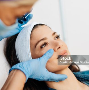
Melasma is a common skin problem, most often seen in women of color. It causes brown to gray-brown patches on the face. Most people get it on their cheeks, bridge of their nose, forehead, chin and above their upper lip. It also can appear on other sun-exposed parts of the body, such as the forearms and neck.

Common melasma triggers include:

  1. Sun exposure: Ultraviolet (UV) light from the sun stimulates the melanocytes. In fact, just a small amount of sun exposure can make melasma return after fading. Sun exposure is why melasma is typically worse in summer. It is also the main reason people with melasma have frequent recurrences.
  2. A change in hormones: Pregnant women often get melasma. When melasma appears in the pregnant patient, it is called chloasma, or the mask of pregnancy. Birth control pills and hormone replacement medicine may also trigger melasma.
  3. Cosmetics: Skin care products that irritate the skin may worsen melasma.

The most important treatment  for melasma is a daily sunscreen that protects against UVA and UVB with an SPF of 30 or higher.  Affected patients should consider alternates to hormonal contraception and irritating skin products should be discontinued. The most popular treatment is with a topical bleaching cream that contains hydroquinone or with compound products that also include a mild corticosteroid and a retinoid. In some patients, chemical peels and laser therapies may be recommended to accelerate improvement. It is important to remember that even minimal exposure to ultraviolet light can cause melasma to recur.

Molluscum Contagiosum

Molluscum Contagiosum

Molluscum contagiosum is a common skin disease caused by a virus that typically presents as a flesh colored bump with a central indentation or dell. This skin disease is most often seen in children and patients who have eczema. Touching something infected with the virus or having direct skin-to-skin contact transmits the virus. Children are more susceptible to molluscum because they often have direct skin-to-skin contact with each other. People who participate in contact sports such as wrestling may also get molluscum from the direct skin-to-skin contact. Teens and adults are exposed to the virus through sexual contact.

Once infected with the virus, a person can spread the virus to other parts the body. Scratching or picking at bumps on the skin and then touching unaffected skin can spread the virus and cause new bumps. The lesions are typically treated with cryosurgery (freezing), curettage (scraping), laser surgery, topical skin therapy, immunomodulators, or retinoids.

Nail Fungus/Nail Lesions

Nail Fungus/Nail Lesions

Nail fungus is also referred to as onychomycosis or tinea ungium. It affects about 12% of all Americans, can run in families and is more common in toenails than in fingernails. Affected nails appear thickened, discolored and may separate from the nail bed, giving the nail a “crumbly” texture . Treatment of fungal infections usually requires oral 2-4 months of oral antifungal medicines. Improvement is very gradual due to the slow re-growth of nails. Topical options are available, but are not as reliable and may take longer to be effective. Most recently, laser procedures have become available as a treatment option. In all treatment modalities, recurrence is very common and occurs in approximately 50% of patients.

Perioral Dermatitis

Perioral Dermatitis

Perioral Dermatitis is a form of rosacea that occurs most commonly in young women between the ages of 20 to 45. Patients present with small red bumps along with redness and peeling around the eyes, nose or mouth. The exact cause is unclear, however the rash appears to be triggered or worsened by one or more of the following: steroid creams, sun exposure and dental products that contain fluoride or cinnamon.

Treatment options include topical creams that have anti-inflammatory properties and oral antibiotics. Most patients improve within one to two months of starting a consistent regiment, however, recurrences are common.

Pityriasis Rosea

Pityriasis Rosea

Pityriasis Rosea (PR) is a very common rash caused by a virus. It is unclear as to how this virus is transmitted, and it does not appear to be contagious. It is most common during the spring and fall.

PR usually begins with a large, scaly pink patch on the trunk called the “herald patch”. Over the course of a few weeks, smaller lesions continue to appear creating what looks like a Christmas tree on the back.

The rash of Pityriasis Rosea may mimic syphilis, a well-known and understood sexually transmitted disease. If a patient has known risk factors for contracting syphilis a blood test will be drawn to rule it out. In most cases a blood test is not needed.

Treatment is usually not necessary as the rash of PR resolves on its own over the course of a few weeks to months with no sequelae. But, topical steroids and treatment with ultraviolet light decreases any itching and hastens resolution in many cases.

Postoperative Instructions

Postoperative Instructions

  1. Leave your bandage on until tomorrow. It is best to remove the bandage at the end of your shower so the adhesive will come off more easily. Always change a bandage if it gets wet from the shower or water exposure.
  2. Gently clean the area with water. If there is stitching, gently dab the stitched area with a Q-tip or gauze with hydrogen peroxide to remove any crusted blood.
  3. All wounds should be gently dried after cleaning with a clean towel or gauze.
  4. Apply a thin layer of ointment to the site. We recommend Aquaphor. If you have tried and used topical antibiotic ointment such as Polysporin, Triple Antibiotic, Bacitracin, or Bactroban and have no history of an allergic reaction in the skin, you may apply any of the above to the area.
  5. If the area is rubbed by clothing during the dayor exposed to irritation, it is recommended to keep it covered using either non-stick gauze (Telfa Gauze) or a simple Band Aid.

(Repeat the above steps till the scab falls off naturally or, if you have stitches in place, until the day of your stitch removal.)

  1. Bleeding may occur after surgery. If you notice bleeding after your procedure, apply firm pressure to the site for 10-15 minutes uninterrupted. If bleeding cannot be controlled then contact your physician at 212-674-0004.
  2. If you notice pus, sudden expanding redness around the wound, or a sudden increase in pain at the site then contact your physician.
  3. It is normal during the first few days to have some swelling and tenderness at the site of the procedure. It is best to take Tylenol (acetaminophen) for any pain unless otherwise advised by a physician.
  4. If you have stitches, avoid excessive motion of the wound area for at least two weeks. Stitch removal is usually painless and is done between 7 to 14 days after the procedure depending on the location and number of stitches. We will make your stitch removal appointment prior to you leaving the office.
  5. Biopsy results are usually available 5 to 7 business days after the procedure. We will call you once they are in the office. If you have not heard after ten days, please feel free to contact us at the office.
  6. Avoid direct sun exposure on the wound for at least six weeks and protect the area with a broad-spectrum sunscreen of at least SPF 30.

If you have any questions, please don’t hesitate to call: 212-674-0004

Psoriasis

Psoriasis

Psoriasis is a chronic, non-curable, common relapsing and remitting immune mediated systemic disease characterized by red, scaly plaques. These skin lesions may vary in severity from localized patches to complete body coverage with nail and joint involvement (psoriatic arthritis).

The etiology of psoriasis is not fully understood. It is generally considered a genetic or inherited disease thought to be triggered or influenced by environmental factors. Psoriasis develops when the immune system mistakes a normal skin cell for a pathogen, and sends out faulty signals that cause overproduction of new skin cells. This over production of skin cells results in the thick, slivery plaque commonly seen. It is not contagious. It tends to flare during the winter. Stress, withdrawal of a systemic corticosteroid, a strep throat infection, and certain medications including beta-blockers, lithium, and anti-seizure medications have each been suggested as a trigger for psoriasis.Injury to the skin can trigger local psoriatic skin changes known as the Koebner phenomenon.

There are a number of treatment options for patients with psoriasis. The decision making process in treating a patient with psoriasis is multi factorial, and depends on the patient’s overall health, presence or absence of joint pain, and severity of the skin involvement. Topical and vitamin D analogues are prescribed in mild cases. In patients with more extensive disease, phototherapy (medically monitored and administered ultraviolet light) may be helpful. More recently, a class of drugs called biologic agents has transformed the treatment of severe psoriasis and psoriatic arthritis. These medications target specific inflammatory molecular markers involved in the disease process. The most commonly used biologics include: Etanercept, Adalimumab, Infliximab, and Usetekinumab. Immunosuppressive medications, like cyclosporine, oral retinoids, and anti-cancer drugs may also be effective in select cases. Newer anti-inflammatory mediations like Apremilast are also gaining momentum.

Restylane ®

Restylane ®

Restylane is a hyaluronic acid injectable gel. Hyaluronic acid is a naturally occurring substance in the skin that adds volume and hydration. With age, our collagen production diminishes and along with it the hyaluronic acid of the skin. Restylane delivers hyaluronic acid to your skin, adding volume and helping to smooth wrinkles and folds.

It is most commonly used for smile lines (also known as nasolabial folds), marionette lines (peri-oral lines), and lip augmentation. The procedure takes approximately 15 to 20 minutes with minimal downtown or recovery and lasts 6 to 12 months.

A topical anesthetic may be used to reduce pain associated with this treatment. Patients are encouraged to arrive 30 minutes prior to their scheduled appointment time to apply the numbing cream.  A mild anesthetic may also be combines with the product to immediately reduce discomfort during the treatment. The discomfort after the procedure is minimal, and Tylenol may be taken for any post-procedural discomfort.

In addition to discomfort, patients may experience minor swelling from the treatment. On average this resolves within 24 hours. However, it may last as long as 72 hours.

We recommend applying ice intermittently after the treatment to reduce swelling. Patients may resume normal activities after receiving treatment, and may apply makeup to conceal any bruising or redness.

Most temporary dermal fillers are composed of hyaluronic acid which is a naturally-occurring chemical predominantly found in skin and connective tissues. Hyaluronic acid is highly effective as a dermal filler because of its ability to transport nutrients to skin.

Additionally, this substance is hydrophilic and plumps facial areas by pulling water to the skin’s surface. Hyaluronic acid is absorbed by the body within six-to-12 months.

Scabies

Scabies

Scabies is an itchy, contagious infestation caused by mites that burrow under the skin. Little red bumps develop in the skin folds, between the fingers, around the belt line and in the genital region. It is transmitted by close personal contact, as well as contact with infested clothing and/or linens.

Treatment consists of either a topical cream called Permethrin or an oral medication called Ivermectin. When prescribed, patients are instructed to apply the cream from the neck down, leaving it on the skin for up to 8 hours. Patients then repeat the treatment one week later. If an oral medication is chosen, patients take one dose on an empty stomach and repeat one week later. Patients are also prescribed a topical steroid to control the itching. Patients are instructed to wash clothing and linens in hot water. Often times, a patient may experience itching or pruritus for up to for up to a month after the initial infestation has been initially treated. Treatment of everyone in the household is required to eliminate the infestation.

Scabies is an itchy, contagious infestation caused by mites that burrow under the skin. Little red bumps develop in the skin folds, between the fingers, around the belt line and in the genital region. It is transmitted by close personal contact, as well as contact with infested clothing and/or linens.

Treatment consists of either a topical cream called Permethrin or an oral medication called Ivermectin. When prescribed, patients are instructed to apply the cream from the neck down, leaving it on the skin for up to 8 hours. Patients then repeat the treatment one week later. If an oral medication is chosen, patients take one dose on an empty stomach and repeat one week later. Patients are also prescribed a topical steroid to control the itching. Patients are instructed to wash clothing and linens in hot water. Often times, a patient may experience itching or pruritus for up to for up to a month after the initial infestation has been initially treated. Treatment of everyone in the household is required to eliminate the infestation.

Seborrheic Dermatitis

Seborrheic Dermatitis

Seborrheic dermatitis is a chronic, relapsing and usually mild inflammatory skin disorder affecting the scalp, face, and torso. Typically, seborrheic dermatitis presents with scaly, flaky, itchy, and red skin. It particularly affects the sebaceous gland-rich areas of skin. In infants, seborrheic dermatitis is called cradle cap and usually resolves in the first year of life. In adolescents and adults, seborrheic dermatitis usually presents as scalp scaling similar to dandruff or as mild to moderate redness of the mid face. Seborrheic dermatitis may occur more commonly in hospitalized patients, those in nursing homes and those with nervous system or immune system diseases.

Mineral oil or gentle shampooing with a mild product can be helpful for children with cradle cap. Seborrheic dermatitis in adults is treated with a topical antifungal or steroid cream, in addition to a medicated shampoo.

Seborrheic Keratoses

Seborrheic Keratoses

Seborrheic Keratoses are benign, non-cancerous, skin growths that may develop anywhere on the skin. They are typically brown or even black in color. They appear pasted or stuck on, are rough in texture, often being mistaken for warts. There may be a genetic predisposition to developing seborrheic keratoses, but they are more commonly associated with maturity. There is no cause for concern and no connection to skin cancer, unless a number of them erupt simultaneously. There are no treatments to prevent seborrheic keratoses from developing, but there are several ways to remove them. They maybe treated with cryosurgery (freezing), curettage (scraping) and/ or electrodessication(lightly burning).

Silhouette Instalift™

Silhouette Instalift™

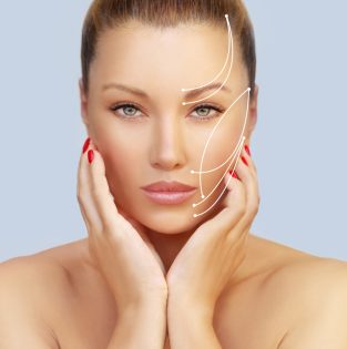
Silhouette InstaLift™ is a non-surgical, in-office procedure that uses completely dissolvable sutures to lift and reposition sagging skin of the face and neck to yield both immediate and long-term results. InstaLift™ uses micro-suspension technology (MST) to provide an immediate lifting effect to the cheeks, jowls, nasolabial folds, jawline, and neck. Over time, InstaLift™ treatment stimulates collagen production to improve skin quality, volume, and thickness.

The Silhouette InstaLift™ is available at GlamDerm to redrape the skin and restore facial structure.

Skin Cancer Screening, Detection, And Treatment

Skin Cancer Screening, Detection, And Treatment

The most important part of our job, as dermatologists, is early detection of skin cancer. We encourage our patients to perform self-examinations frequently, bring suspicious growths to our attention and come in for a full body skin examination annually. In select cases, where patients have a number of melanocytic nevi or brown moles and/or a familial history of malignant melanoma, we may recommend total body examinations twice a year.

Skin Tags

Skin Tags

Skin tags are benign, skin colored or brown growths that are most commonly located on the neck, the armpit or in the groin area. The cause of skin tags is thought to be friction. They are more common in people who are overweight or who have diabetes. Treatment is not necessary, but removal for cosmetic purposes can be accomplished with surgery, cryosurgery (freezing) or electrodessication (burning).

Squamous Cell Carcinoma

Squamous Cell Carcinoma

After basal cell carcinoma, squamous cell carcinoma is the second most common type of skin cancer. Middle aged or elderly fair skinned individuals who have significant sun damage are most affected by this form of skin cancer. Patients, who have undergone organ transplantation, have chronic ulcers, a smoking history, history of toxic exposures, and immunosuppression are considered high risk.

Actinic keratoses or precancerous lesions often precede squamous cell carcinomas. Clinically, squamous cell carcinomas, appear scaly, crusted and inflamed. They may grow rapidly, and often ulcerate or bleed spontaneously. These lesions, typically, occur on sun-exposed body parts like the head, neck, face, ears or backs of hands. Squamous cell carcinomas may also affect the lips, oral cavity or on the genitalia.

Given their potential to grow, locally invade nearby tissue, and spread to distant body parts (metastasize), early intervention is important. Patients require a thorough evaluation and then a skin biopsy to confirm the diagnosis. Once the diagnosis is confirmed by a dermatopathologist after looking at the specimen microscopically, a variety of treatment options are available depending on the individual patient, as well as the size and location of the tumor. These options include an in office surgical excision under local anesthesia, Mohs surgery, cryosurgery (freezing), radiation, electrodessication and curettage and topical chemotherapeutics.

Sun Protection/Sunscreen

Sun Protection/Sunscreen

Based on their mechanism of action, topical sunscreens can be broadly classified into two groups, chemical absorbers and physical blockers. Chemical absorbers work by absorbing ultraviolet (UV) radiation and can be further differentiated by the type of radiation they absorb, UVA or UVB, or both UVA and UVB. Physical blockers work by reflecting or scattering the UV radiation. The most common physical blockers are titanium dioxide and zinc oxide.

Sunscreen can protect your skin against skin cancer and premature aging. However, it is not as effective unless it’s applied correctly. Follow these tips from dermatologists when applying sunscreen:

  1. Choose sunscreen that has an SPF of 30 or higher, is water resistant, and provides broad-spectrum coverage, which means it protects you from UVA and UVB rays.
  2. Apply sunscreen generously before going outdoors. It takes approximately 15 minutes for your skin to absorb the sunscreen and protect you. If you wait until you are in the sun to apply sunscreen, your skin is unprotected and can burn.
  3. Use enough sunscreen. Most adults need at least one ounce of sunscreen, about the amount you can hold in your palm, to fully cover all exposed areas of your body. Rub the sunscreen thoroughly into your skin.
  4. Apply sunscreen to all bare skin. Remember your neck, face, ears, tops of your feet and legs. For hard-to-reach areas like your back, ask someone to help you or use a spray sunscreen. If you have thinning hair, either apply sunscreen to your scalp or wear a wide-brimmed hat. To protect your lips, apply a lip balm with a SPF of at least 15.
  5. Reapply sunscreen at least every two hours to remain protected, or immediately after swimming or excessively sweating.

People who get sunburned usually didn’t use enough sunscreen, didn’t reapply it after being in the sun, or used an expired product.

Your skin is exposed to the sun’s harmful UV rays every time you go outside, even on cloudy days and in the winter. So whether you are on vacation or taking a brisk fall walk in your neighborhood, remember to use sunscreen.

Surgical Procedures – Post Op

Surgical Procedures – Post Op

What to arrange in advance:

  • Post operatively you may need to limit your range of motion in the area of the surgery to minimize risk of stretching the scar or causing stitches to pop out. Please plan accordingly.
  • After your surgery, you will need to change and dress the site daily. Make arrangements so that you can have help if it is a difficult to reach area.
  • If your wound requires sutures, then you will need to return to our office for suture removal 5-14 days later depending on the body location. Plan your surgery at a time where you can return back to the office in the recommended amount of time. Make your suture removal appointment with the front desk on the day of your surgery.
  • There may be lab fees associated with your surgery. An outside laboratory, not GlamDerm bills these fees. Please check with your insurance company in advance of your surgery to ask if lab fees are covered.

If you have any questions, please call the office 212-674-0004.

Surgical Procedures – Pre Op

Surgical Procedures – Pre Op

Prior to your surgical procedure, please find a list of our recommendations below.

  1. Avoid alcohol consumption the week prior to your procedure.
  2. Stop all alternative, homeopathic treatments including garlic, ginkgo, licorice extracts, fish oil and vitamin E doses over 400 units one week before surgery. It is unknown what effect these substances may have on bleeding and wound healing.
  3. Stop pain medications including aspirin, Advil, Motrin, Ibuprofen, Aleve, etc. for one week prior to the procedure. Tylenol is ok to take, as it does not increase the risk of bleeding.
    Please note: If aspirin is taken for a prior history of stroke or cardiac events, check with your appropriate doctor to see if it may be prior to surgery.
  4. Aspirin, Advil, Motrin, Ibuprofen, Aleve may be resumed 2 days after surgery if no unusual bleeding has occurred.
Telangiectasias

Telangiectasias

Telangiectasias, or broken capillaries, are small linear vessels that may appear on the face, around the nose or the legs. They maybe part of an inherited condition, acquired later in life, or secondary to certain the use of medications like topical steroids. These lesions are generally harmless. Treatment is usually sought secondary to aesthetic concerns. Telangiectasias may be treated by intense pulse light (IPL), vascular laser treatment, electrosurgery (burning) or sclerotherapy.

Threadlift

Threadlifts Are the New Trend

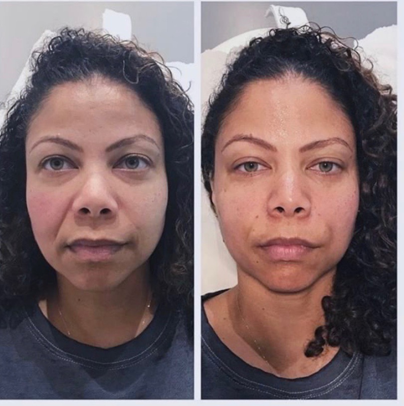
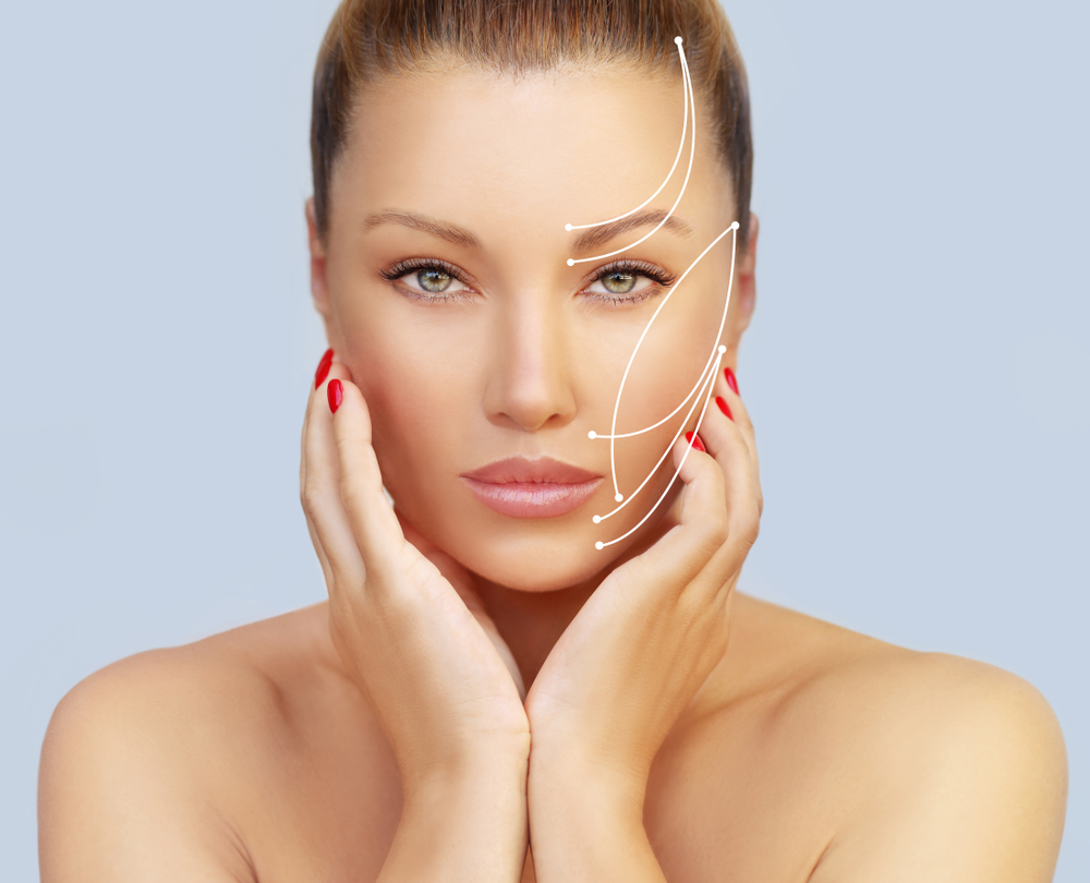
Skin laxity can present in varying degrees. However, when women and men begin to notice falling cheeks, developing jowls, and a sagging neck, many decide it’s time for facial rejuvenation.

That being said, a traditional facelift can cost upwards of $10,000 and require weeks of recovery.

Fortunately, a threadlift can provide patients with a non-surgical and more cost-effective alternative, with minimal downtime. With so many advantages, it’s not surprising that threadlifts are the new trend to look younger and refreshed without surgery.

So, is a threadlift right for you? Keep reading to learn all about the new and improved threadlift and what patients can expect should they decide to undergo this procedure.

Tinea Versicolor

Tinea Versicolor

Tinea versicolor manifests itself clinically as small, scaly, white, pink or brown spots on the upper arms, chest, back, neck or face. The rash is most noticeable in the summer and on dark skinned patients because the causative organism prevents normal tanning of the skin.

Tinea versicolor is caused by an overgrowth of yeast on the skin. The yeast favors the pores of oily skin and grows best in hot, humid weather. This infection is most common in teenagers and young adults, but can affect any age. A diagnosis of tinea versicolor is usually based on its clinical appearance. In some cases, a skin scraping will be done or a special light, called a Wood’s lamp will be used to confirm the diagnosis.

Topical antifungal creams and in some instance oral medications are the mainstays of treatment. Shampoos that contain selenium sulfide or pyrithione zinc may also be recommended for use on the body. It is important to remember that although the organism is easy to eradicate, it can take weeks to months to regain normal pigmentation.

Vitiligo

Vitiligo

Vitiligo presents as symmetric milky, white patches, most commonly on the face, lips, hands, arms, legs and genitals. Half of those affected are under the age of 20, and 1 in 5 have affected family members.

Vitiligo is believed to be an autoimmune process. Autoimmune disease develops when self attacks self. In the setting of vitiligo, the body attacks its own pigment producing cells, the melanocytes. Most patients with vitiligo are otherwise healthy. Occasionally, vitiligo may occur with other autoimmune diseases like thyroid disease or alopecia areata.

The course and severity of pigment loss varies from patient to patient. There is no way to predict how much color an individual will lose. In general, because of the contrast in color, vitiligo appears more obvious in patients with skin of color.

Treatment of vitiligo begins with topical corticosteroids and calcineurin inhibitors, like pimecrolimus and tacrolimus. Other options include light and laser therapy. In extensive disease, where repigmentation is not practical, complete chemical depigmentation may be considered.

Vollure

Vollure

Juvederm Vollure is a Hyaluronic Acid for correction of moderate to severe facial wrinkles and folds, such as nasolabial folds that lasts up to 18 months. Nasolabial folds are static lines that appear around your nose and mouth when your face is at rest.

This happens over time as your skin loses some of the building blocks (hyaluronic acid and collagen) that give the skin its structure. Vollure maintains correction and also allows easy movement in the treated area with a molecular weight mix specific for this area.


Voluma XC ™

Voluma XC ™

Juvederm Voluma XC is the first and only hylauronic acid filler that is FDA approved to instantly add volume to the cheek area, resulting in a subtle lift. It has been clinically proven to last up to two years with optimal treatment.

A topical anesthetic may be used to reduce pain associated with this treatment. Patients are encouraged to arrive 30 minutes prior to their scheduled appointment time to apply the numbing cream. Voluma contains an anesthetic allowing patients to feel more comfortable as the treatment progresses. The discomfort after the procedure is minimal, and Tylenol may be taken for any post-procedural discomfort.

In addition to discomfort, patients may experience swelling from the treatment. On average this resolves within 24 hours. However, it may last as long as 2 weeks in some rare instances. We recommend applying ice intermittently after the treatment to reduce swelling. Patients may resume normal activities after receiving treatment, and may apply makeup to conceal any bruising or redness.

Warts

Warts

Warts are common, non-cancerous growths caused by an infection with human papilloma virus. There are several different types of wart and their appearance varies depending on their location.

Plantar warts are located on the soles of your feet, while palmar warts grow on the palms of your hands. In both locations, these warts appear pushed in either from pressure of hand use or pressure from walking. They are flat with tiny black dots and loss of the skin lines. Plantar warts may feel uncomfortable, and are often times painful.

Common warts affect the fingers, around the nails and on the backs of the hands. They are raised and skin colored with a rough surface and black dots.

Flat warts are smaller and smoother in texture. They grow in clusters, and occur predominantly on the face.

Genital warts, medically known as condyloma accuminata, are sexually transmitted. They are flesh colored, may be smooth or rough, and can appear on any area of the genital region including inside the vagina, on the cervix, on the anal verge and within the rectum. In women, some strains of genital warts are associated with an increased risk of cervical cancer while, in men, perianal
warts may carry a risk of squamous cell carcinoma. Careful follow up with a gynecologist or a proctologist, respectively, is necessary in these cases.

There are a variety of treatment modalities available to eradicate warts. Cryosurgery (freezing) is most commonly used to destroy warts, but electrosurgery (burning) or an application of an acid may also be done. An alternative approach is immunotherapy, in which the body’s immune system is triggered to fight the wart using a variety of agents (e.g. imiquimod, cantharidin, candida antigen). Additional options include injectable chemotherapeutics (e.g. bleomycin) and other medications that prevent the wart virus from dividing (e.g. podophyllin). Regardless of the treatment used, repeat treatments are usually required.

Xeomin ®

Xeomin ®

Neurotoxins are purified proteins that have been used for years to treat dynamic wrinkles that form from excessive small muscle contraction. These proteins have been used to smooth and relax frown lines, crow’s feet, forehead wrinkles, upper lip wrinkles, neckbands, and “gummy smiles.”

These toxins are highly purified and dilute, and have proven to be extremely safe to use. The toxin is injected directly into the desired area and takes effect by decreasing the contraction of the muscle, ultimately softening and in some instances eliminating the line. Relaxation is gradual, usually starting after 48 hours with Dysport and 72 hours with Botox, progressing over 2 weeks. The treatment lasts on average for three and a half to four months.